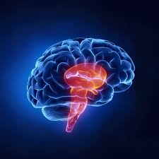

The neurons in the dorsal cortex receive visual and sensory inputs from the thalamus on apical dendrites that expand across the cortical surface. In addition, it was not thought to be more extensive than the dorsal cortex of egg-laying vertebrates.Įarly works identified similarities between the patterns of connection between neuronal types in the dorsal cortex of reptiles and the neurons of the neocortex. The dorsal cortex makes up a small region of the forebrain and contains a thin layer of pyramidal neurons and inhibitory interneurons.įrom the observation of fossil records, it is thought that early mammals had a small neocortex relative to the size of the olfactory bulb and appeared as a small cap at the top of the forebrain. A large amount of research investigating the neocortex’s origin focuses on similarities between the dorsal cortex and the neocortex. The Neocortex and the Dorsal Cortexĭespite intense research spanning over a century, the evolutionary origin of the neocortex is still unclear. Among its functions are to process sensory information and derive language, emotions, and meaningful memories.Īdditionally, it is responsible for declarative memory, which is memory that can be spoken aloud (such as learned facts), and is further divided into two subgroups- semantic and episodic memory. The temporal lobe houses the hippocampus and the amygdala. It hosts the primary area for visual perception which is closely surrounded by the visual association area. The occipital lobe is responsible for visual function and is the bulge seen at the back of the brain. Traditionally thought as the association cortex, the parietal lobe is believed to play a role in decision-making, numerical cognition, processing of sensory information, and spatial awareness. Disorders of the frontal lobe include frontotemporal dementia, Parkinson’s disease, and Alzheimer’s disease. In this region of the neocortex is the human executive function that manages the intricacies of multiple complex processes such as task switching, reinforcement learning, and decision-making to name a few. The frontal lobes are responsible for the selection and coordination of goal-directed behavior. Research on brain function shows that each neocortical layer has specific functions. The neocortex is comprised of 4 regions based on the patterns of sulci (grooves) and gyri (ridges) in the brain: frontal, parietal, occipital, and temporal lobes.

Role of the transketolase-like 1 gene in the development of the modern human brain.Beta-amyloid deposits found in young patients with COVID-19.New insights into the understanding of how ApoE variants contribute to Tau pathology in Alzheimer’s disease.Molecular later: contains a very small number of neurons,Įach layer is characterized by varying neuronal shapes, density, sizes, and organization of nerve fibers.In order of superficial to deep, the layers are Roman numerals are used to number the six neocortex layers. In just one hemisphere of the neocortex, there are an estimated 8 billion neurons.
#Cerebro reptiliano plus#
It is comprised of six layers of neuronal types: one layer of axon endings and apical dendrites from cortical neurons plus five cell layers. The neocortex itself is essentially like a sheet of tissue that varies in thickness but has a very large surface area. Some cortical areas have been extremely well described, whereas others pose more puzzling and disagreement over function exists.

Scientists divide the neocortex into smaller cortical areas that are often referred to as the “organs of the brain”.

The human brain differs from brains of other animals and even mammals based on its relatively larger size and predominance of the neocortex. Lecture 3: The Structure of the Neocortex Play Structure of the Neocortex


 0 kommentar(er)
